|
» »
Exercises for herniated disks, protrusions, and osteochondrosis
Author: Pavel Shaikin - master trainer in fitness (FPA). All articles by the author ➤
TABLE OF CONTENTS:
This issue is relevant among practitioners. According to WHO statistics, 80% of the population suffers from diseases of the musculoskeletal system - spine, joints, ligaments, and muscles, particularly those of working age (30 to 50 years). Sometimes osteochondrosis manifests even in adolescence and continues to progress thereafter. The term "osteochondrosis of the spine" refers to a primary degenerative process in the intervertebral discs, which in turn leads to secondary reactive and compensatory changes in the bony-ligamentous apparatus of the entire spine. Osteochondrosis of the spine is a civilization disease associated with bipedalism and the conditions of its exploitation. 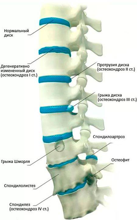
It can be asymptomatic or can occur against a backdrop of various symptoms that hinder activity or a fulfilling life. Generally, people become interested in spine health issues only when symptoms appear, which is not quite right. Initial prevention is necessary to avoid disease progression, even for individuals without signs of osteochondrosis. Such prevention is already "embedded" in the theory and methodology of fitness activities. A solid foundation in training is essential, allowing for the simultaneous preservation or improvement of health and achieving desired results. The foundation for spinal health is a well-developed muscle corset of the spine, good intermuscular coordination, and correct exercise technique. It seems clear that one needs to strengthen muscles and adhere to proper exercise techniques. However, there lies a misleading path that may lead to the exacerbation of osteochondrosis and its symptoms in the future. The problem lies in the misinformation from various sources on these matters. Individuals who already have intervertebral hernias and protrusions also encounter these sources while searching for various exercises and training for osteochondrosis. Basics of Anatomy and Physiology of the Spine, OsteochondrosisLet's delve into the anatomy of the spine and physiology to clarify the overall picture and to serve as a foundation for addressing any misunderstandings that may arise later. The spine consists of the bodies of the vertebrae, which are separated by intervertebral discs (IVD). They resemble an onion with layers, consisting of a pulpy nucleus and a fibrous ring. The pulpy nucleus is a gelatinous fluid that can extend beyond the boundaries of the fibrous ring, leading to the formation of intervertebral hernias (IVH). 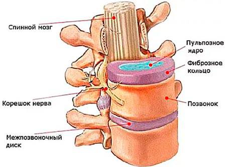
The function of IVDs is to cushion the vertebrae. Together with various ligaments of the spine and deep muscles, the vertebrae are stabilized relative to each other. The deep muscle layers contain blood vessels and supply the spine with fluid and nutrients – this is the primary source of nutrition for the IVDs after the age of 18-23 years. The anatomical curves of the spinal column have protective functions, provide support for the body, and assist in locomotion (movement), while also working in conjunction with the pelvis and lower limbs for amortization. 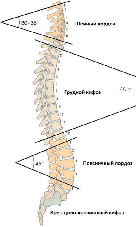
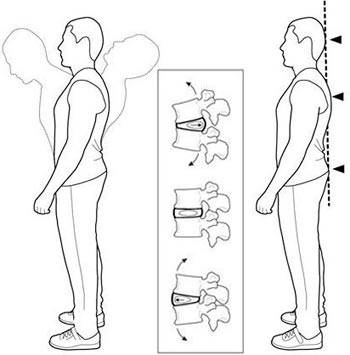
The cause of protrusion and hernia formation is uneven loading on the spinal column, specifically increased pressure between two vertebral bodies on the IVD at an angle. The position of the spine that deviates from its natural neutral position places compressive load at an angle on the spinal segments. (The neutral position of the spine (NPP) is the state of the musculoskeletal system in which a neutral zone is maintained across all segments of the spine. In the neutral position, the safest position for the spinal joints is provided, especially for the intervertebral discs). When this compression is maintained for an extended period, degenerative-dystrophic disorders of the cartilage and muscles develop, causing dehydration of the IVDs, which leads to flattening of the vertebral bodies (decreasing the distance between them). Simultaneously, some spinal ligaments become overstretched, potentially causing spinal instability and thickening of the ligaments. 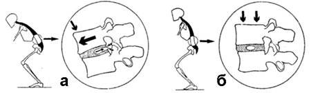
A - forces acting on the IVD displace the pulpy nucleus toward the spinal canal from a bent position of the spine, where the vertebral bodies form an angle. B - load is evenly distributed across the IVD, and the pulpy nucleus is centered. From the perspective of the muscular system, the deep muscles of the spine attempt to return the spine to its neutral position while constantly experiencing tension. This tension compresses the vessels that nourish them, disrupting circulation (ischemia). Muscle fibers gradually atrophy, turning into connective tissue. The connective tissue then fails to nourish the IVDs, leading to the progression of osteochondrosis, potentially resulting in the formation of osteophytes and ossification of the vertebral bodies, culminating in a complete loss of mobility in advanced cases. Uneven loading on the spine over extended periods arises from functional deficiencies in certain muscles and postural disorders, such as scoliosis. There are many causes for postural violations, one of which is obesity and unbalanced load on various muscle groups during training. The clinical manifestations of osteochondrosis are highly diverse. Based on localization, spinal osteochondrosis is classified as cervical, thoracic, or lumbar. There are numerous neuralgic syndromes associated with osteochondrosis of the spine, including root syndrome, shoulder-scapular arthritis, cardiac syndrome, vertebral artery syndrome, radiculitis, lumbago, sciatica, and intercostal neuralgia. Root syndrome is the most commonly encountered. Various symptoms of osteochondrosis (headaches, muscle pains, tingling, coldness, itching, burning, cramps, general weakness, etc.) may manifest when there is irritation or compression of the nerve roots of the spinal cord. The localization of pain depends on which roots are irritated by protrusions, IVHs, or bony growths (osteophytes). 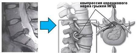
In terms of root syndrome, the muscles, influenced by mechanical overloads and pathological impulses from the affected spinal motion segment (SMS), develop a persistent contracture, which disrupts the mobility of the vertebrae and circulation, exacerbating postural disorders, creating a vicious cycle. 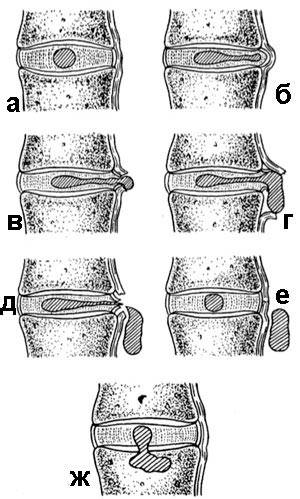
Various stages of intervertebral disc herniation formation (according to Arseniy K. et al., 1973)
a - normal intervertebral disc. The main goal of training is to halt the progression of osteochondrosis and restore normal functions to the muscles that will nourish the IVD, dissolve the IVH, and stabilize the spine. Additionally, enhancing the functional capabilities of the body will help partially compensate for various limitations associated with osteochondrosis of the spine. Causes, Treatment, and Prevention of Osteochondrosis within FitnessThe technology of fitness training is designed for body health improvement and prevention of various diseases, while simultaneously addressing various goals of the practitioners. Therefore, proper fitness training improves the situation as a whole. When dealing with osteochondrosis of varying stages and segments, there are specific considerations for gym work. Let's discuss general recommendations for the training process. 1. Exercise technique should be correct, regardless of the level of fitness and the presence/absence of osteochondrosis. In the case of protrusions and IVHs, exercises with incorrect technique increase their sizes, especially when compression of the vertebrae occurs at an angle. Exercises should be as natural as possible to reduce the risk of injury. 2. Training should focus on correcting postural abnormalities and scoliosis, regardless of the presence or absence of osteochondrosis. Postural abnormalities can take many forms in both the sagittal and frontal planes, and in the case of scoliosis, they affect the frontal and horizontal planes. Based on these abnormalities, exercises and their dosage in terms of volume and intensity should be selected. 3. The load should be balanced across antagonist muscles (equal sets and resistance for major opposing muscle groups), regardless of the presence or absence of osteochondrosis considering postural changes. An unbalanced load leads to increasing discomfort in the comfortable exercises, resulting in greater muscle imbalance, while also increasing stress on joints and muscles that must adapt to the new position of the bones. Strong muscles may shorten, while stretched ones weaken, disrupting blood circulation, leading to muscle myofibrils (contractile protein structures) turning into connective tissue (fascia). 4. Excess body weight and obesity increase the risks of problems with the musculoskeletal system, including causing postural abnormalities and uneven loading on the spine. It is essential to maintain a normal body fat percentage. 5. A weak or non-functional muscle corset limits the ability to perform exercises with correct technique. Often, this refers to a non-functional spinal corset or non-functional shoulder blades. It is necessary to develop specific physical functions in the muscles rather than just bulk them up. 6. Insufficient mobility in joints can lead to excessive mobility in spinal joints, exacerbating the manifestation and progression of osteochondrosis. It is important to identify joints with insufficient mobility and increase it. 7. Excessive intensity of training leads to under-recovery of the musculoskeletal system. Accumulated under-recovery over an extended period leads to wear of the joints, increasing the risks of osteochondrosis. The load should follow a periodization model to allow all systems of the body to recover. At present, there is a variety of information regarding all 7 points, including conflicting information. Here lies the problem of misinformation, and there is so little accurate information that it is often overlooked. Given the principle of individual differences, no article can reveal specific actions in dealing with osteochondrosis – there are too many variables, from posture and mobility to segmental muscle and IVD disorders. Failure to adhere to these recommendations limits your physical development and training results. How I Address Prevention of Progression of Osteochondrosis, Protrusions, and IVH in My Workouts:1. Assessment of posture and joint mobility. Correction of postural deviations, improvement of mobility in restricted joints, and reduction of excess body weight in cases of obesity. 2. Possible alleviation of muscle spasm, swelling, and pain through specific movements (somatic correction, PNF), during which proprioceptors send signals to the CNS, leading to the relaxation of spasmed muscles and improved circulation. 3. Establishing a neutral spine position in many exercises. Initially, I use a gymnastics stick and a mechanical tonometer to monitor neutrality. Later, I transfer this awareness to more complex exercises and everyday life. The neutral position is necessary to avoid exacerbating protrusions and hernias. 4. Development of muscular endurance to enhance blood circulation, elasticity, and nutrition for the spine through fluid diffusion. This includes certain exercises from kinesitherapy that do not cause compression of the IVDs and pain. Muscle engagement triggers self-recovery mechanisms. 5. Development of various functional qualities of the muscles, including rhythmic stabilization on unstable surfaces to engage the deep spinal muscles. 6. Therapeutic methods to improve tissue nutrition and remove waste products – contrasting showers with stretching of specific muscles, massage, and work with myofascial pain syndromes. 7. Healthy and adequate nutrition. As you can see, a comprehensive strategy is needed to address the condition, not just isolated exercises from kinesitherapy or muscle strengthening that could be harmful. Common Mistakes When Training with OsteochondrosisThe following information is more specific and will assist you in your training. I want to note that it may contradict established stereotypes: 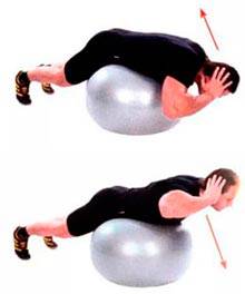
- Hyperextensions with a rounded back. This technique contributes to spinal compression and the extrusion of the pulpy nucleus into the spinal canal. The solution is to either exclude the exercise or maintain a neutral spine position while carefully working on the spinal extensors with a stability ball. - Strengthening the back, including kinesitherapy. It is essential to understand postural abnormalities and where to strengthen muscles rather than overloading those that are already capable of compressing vertebrae at an angle and disrupting circulation. The solution is to consult a competent trainer. - Strengthening superficial muscles - an incorrect strategy for combating osteochondrosis. The solution is to focus on developing specific deep spinal muscles on unstable supports and practicing maintaining a neutral spine. - It reduces the load on the spine. A significant portion of spinal injuries is sustained here! In reality, the load on the spine is substantial (due to stabilizing muscles), which creates compression in the lumbar region and can cause muscle strains or misalignment of the vertebrae under heavy loads. The solution is to minimize the weights or, if bodyweight squats are insufficient, replace them with single-leg squats using TRX or step-ups. These exercises exclude the load on the spine. That’s true if the exercise technique is correct. This is often violated when the pelvis tilts relative to the spine. The solution is to reduce the weight and monitor the position of the pelvis. Here, the neutral position of the spine is disrupted, which may increase the progression of protrusions and hernias. The solution is to maintain the spine in a neutral position. - Exercises are safe without axial load on the spine. When the neutral position of the spine and exercise technique are compromised, any exercises can become dangerous even without axial load. Here, the neutral position of the spine is also disrupted, leading to hyperlordosis and compression of the IVDs. The solution is to keep the spine neutral while slightly engaging the abdominal muscles. 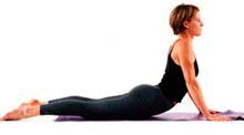
- The Cobra Pose from yoga for stretching the spine and back muscles. In fact, the spine and back muscles move closer together, compressing the IVDs. The solution is to exclude this exercise and similar ones. For improving mobility, gently use the Cat-Cow stretch from a quadruped position. One of the working muscles is the iliopsoas, which in this exercise compresses the IVDs, shifting the bodies of the vertebrae forward. The solution is to include alternate leg raises from a lying position on your back at certain stages. 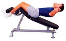
- Roman chair for abs. The situation is the same as with leg raises, only there is more compression. The solution is the same as with leg raises. - Leg swings exclude load on the spine. The technique in leg swings is often incorrect, causing the pelvis to tilt relative to the spine, thereby creating compression on the IVDs. The solution is to either exclude the exercise or carefully monitor the position of the pelvis and spine, which can be very challenging. - Treatment of the spine involves exercising through pain. This concept originated in kinesitherapy. Exercising through pain can lead to increased hernias and protrusions when compression of the IVDs occurs under pressure. The solution is to avoid exercises that cause persistent pain and compression of the IVDs. These exercises often overload the lumbar area due to the bridge position and unstable supports at five points. The solution is to maintain all five points of support (feet, sacrum, shoulder blades, and back of the head) and keep the spine in a neutral position. The feet can be placed wider than shoulder-width on the steps to reduce the bridge effect. - The absence of pain during exercise is an indicator of a suitable exercise and correct technique. This is a dangerous misconception. With compression of the IVDs, the appearance of protrusions, or increased IVH, there may be no pain as the IVDs lack pain receptors, while pain arises from irritation of the nerve roots. Comfortable exercises can increase muscle development imbalances, leading to unequally distributed loads on the joints. The solution is that exercises should be natural and performed with correct technique. - Aerobic exercise improves blood circulation in the muscles and contributes to the treatment of osteochondrosis. It can be said that aerobic exercise improves overall cardiovascular health, but it cannot be said to improve blood circulation in the deep spinal muscles. The solution is to include exercises with moderate or light resistance in a gym setting. - Diets for weight loss and fasting. In attempts to reduce body weight, people seek out ineffective and even harmful diets, after which weight often returns within a few months or years. The solution is a comprehensive approach to fat burning and healthy eating. - Hanging on the bar for spinal stretching. This can overstretch the spinal ligaments, increasing instability, and afterwards, compression of the vertebrae occurs due to the contraction of the deep muscles, worsening the situation overall. The solution is to avoid hanging and stretch from a lying position on your back, extending through the top of your head and arms. In some cases, you can stretch using a modified hang where there is support for the feet, followed by an equal amount of time lying on your back in a horizontal position. - With IVH in the cervical spine, it is essential to strengthen the neck muscles. Typically, neck pain and weakened neck muscles are compensations for dysfunctions lower down the kinetic chain. In these cases, strengthening the neck will worsen the situation. The solution is to identify postural and mobility issues in the joints below the neck and correct any identified deviations. This list of mistakes could go on for a long time. Most of these errors relate to disruption of the neutral spine position, stretching of the muscles and spine, attempts to bulk up the back muscles, and a lack of reliable information about your posture and joint mobility for setting correct training goals. The conclusion is this – do not use a trial-and-error method in your training; it is too lengthy a path that can harm your health. To train with osteochondrosis and its progression effectively, it is essential to work under the guidance of a competent professional if your health, time, and training results (weight loss, muscle gain, posture correction, etc.) are important to you. Train wisely! Contacts for Pavel: VK, Facebook, Website. SIMILAR ARTICLES
COMMENTS |