|
»
Sports shoulder injuries: types, symptoms, treatment
Author: Sergey Yezhov - sports medicine physician. All articles by the author ➤
I have already discussed knee injuries on the site here. Today, we will talk about the shoulder joint, which is similarly complex in structure and functionally overloaded. I want to clarify that I will not describe superficial injuries such as bruises and sprains. Instead, let’s focus on more specific injuries related to the shoulder. It’s worth mentioning the anatomical features of the shoulder joint. It is formed by the head of the humerus and the acromial cavity of the scapula. The joint lacks supporting and stabilizing ligaments. Therefore, the humeral head is essentially suspended by the joint capsule and stabilizing muscles. This provides a large range of motion in the joint but also increases the risk of injury. Additionally, there is a labrum made of cartilage along the edge of the acromial cavity. This can be damaged during impact with the humeral head, leading to chronic pain in the joint. Injury to the Rotator Cuff Tendons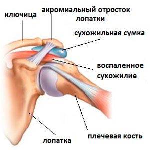
This is a relatively common injury among athletes. As I mentioned earlier, the humerus is effectively suspended by the muscles. These include the subscapularis, infraspinatus, supraspinatus, as well as the teres minor and major muscles of the scapula, the deltoid muscle, and the coracobrachialis. This anatomical feature increases the risk of injury, as well as chronic static overload of the joint during sports activities. Acute injury typically occurs due to a sudden twisting of the arm or from falling on an outstretched arm. Clinically, it presents as pain in the area of the shoulder, which intensifies with abduction and rotation of the arm. There is usually no significant swelling of tissues or other visible pathology. A precise diagnosis is made based on the results of ultrasound or MRI of the shoulder joint. In the case of a complete tear of one or more tendons, surgical intervention is required. In the event of a partial tendon injury, the arm is immobilized in a sling for up to 7-10 days. Non-steroidal anti-inflammatory drugs (NSAIDs) are prescribed both orally and topically in the form of ointments, along with physiotherapy and therapeutic exercise. Sports activities should not be resumed for at least 3 weeks after the injury. With prolonged, chronic overload, a condition known as subacromial impingement syndrome can develop. This is inflammation of the supraspinatus tendon, which gets compressed by the acromial process of the scapula. This syndrome more often occurs in sports that involve frequent overhead arm movements (volleyball, artistic gymnastics, tennis, swimming). It is also commonly seen among amateur strength training enthusiasts. It is characterized by sharp pain when raising the arm above 90 degrees. There may also be a "clicking" sensation in the joint. Treatment of this condition should be carried out under strict medical supervision. As a rule, anti-inflammatory medications, neuro vitamins, massage, balneotherapy, and physiotherapy are prescribed. In severe cases, surgical treatment may be necessary. Returning to sports should be gradual, and it’s advisable to work with a qualified coach during the initial period. Shoulder DislocationThe primary causes of shoulder dislocation can include direct blows to the shoulder joint area, falls onto an outstretched arm, or rotational arm movements involving force. However, shoulder dislocation presents a significant problem during constant strength training; it can recur repeatedly during presses, pull-ups, and other exercises that engage the shoulder joint. When diagnosed with a shoulder dislocation, the symptoms may include:
Treatment for a shoulder dislocation occurs in several sequential stages. First and foremost, first aid should be provided. If you are not a doctor, do not attempt to manipulate the injured area; it is best to call for and wait for emergency medical help or transport the individual directly to the hospital. First aid for a shoulder dislocation that can be performed before a doctor's examination includes:
After confirmation of the diagnosis, treatment is prescribed based on the severity of the injury. In any case, the reduction of the dislocation should only be performed by a physician. Sometimes, during a shoulder dislocation, there can be extensive damage to the joint capsule. This leads to chronic instability and the development of habitual dislocations. In such cases, I recommend strengthening the shoulder muscles using a special figure-eight supporting bandage. If this method proves ineffective, surgical treatment may be necessary. At the end of this article, I would like to discuss another common injury. While it's not directly related to the shoulder joint, it is highly relevant for enthusiasts of strength sports. Injury of the Pectoralis Major MuscleTypically, a tear occurs due to excessive tension in the muscle. For example, during the performance of This is a common cause of tears in about half of the cases, or from a blow to the arm at the moment when the pectoralis major muscle is tensed (in volleyball, with arm wrestlers, during rugby, etc.). There are 5 types of tears in the pectoralis major muscle:
At the moment of injury, the affected person may sometimes hear a cracking sound. Immediately afterward, pain appears in the chest or shoulder area. The pain may "shoot" down the arm. Within a few minutes, a bruise may appear on the shoulder, which is a classic indicator of a tear. Over the next few days, the bruise can spread to the upper limb or abdomen. In the case of a complete rupture, the muscle retracts and pulls towards the sternum, and a depression becomes visible at the site of the rupture. Since the torn muscle cannot fully perform its function, weakness arises in movements for which this large muscle is responsible (pushing from the chest, bringing the arm towards the body, etc.). Later, as the acute pain from the tear decreases, strength gradually recovers, but with the compensation of other muscles. Unfortunately, in the case of a complete tear, self-recovery of strength is never complete: without surgery following a complete tear, strength only recovers to about half! In partial tears, there is no depression, and bruising may be small or entirely absent. This creates certain challenges for diagnosis. A correct diagnosis is made based on the results of ultrasound or MRI of the thoracic muscle. In the case of partial damage to the pectoralis major (up to 30% of fibers), it is essential to ensure complete rest of the limb by suspending it in a sling. Anti-inflammatory ointments are applied locally. From the second week post-injury, physiotherapy procedures (magnetotherapy, UHF) are prescribed. Gym activities can resume no earlier than 6 weeks after injury. In cases of more severe damage to the pectoralis major muscle, surgical treatment is employed: the damaged area is sutured. Returning to training is possible no earlier than 6 months post-surgery. SIMILAR ARTICLES
COMMENTS |